Chapter 7: Structural Organisation in Animals
In the preceding chapters you came across a large variety of organisms, both unicellular and multicellular, of the animal kingdom. In unicellular organisms, all functions like digestion, respiration and reproduction are performed by a single cell. In the complex body of multicellular animals the same basic functions are carried out by different groups of cells in a well organised manner.
A group of similar cells along with intercellular substances perform a specific function. Such an organisation is called tissue. You may be surprised to know that all complex animals consist of only four basic types of tissues.
Animal Tissues
The structure of the cells vary according to their function. Therefore, the tissues are different and are broadly classified into four types:
- Epithelial Tissue
- Connective Tissue
- Muscular Tissue
- Neural Tissue
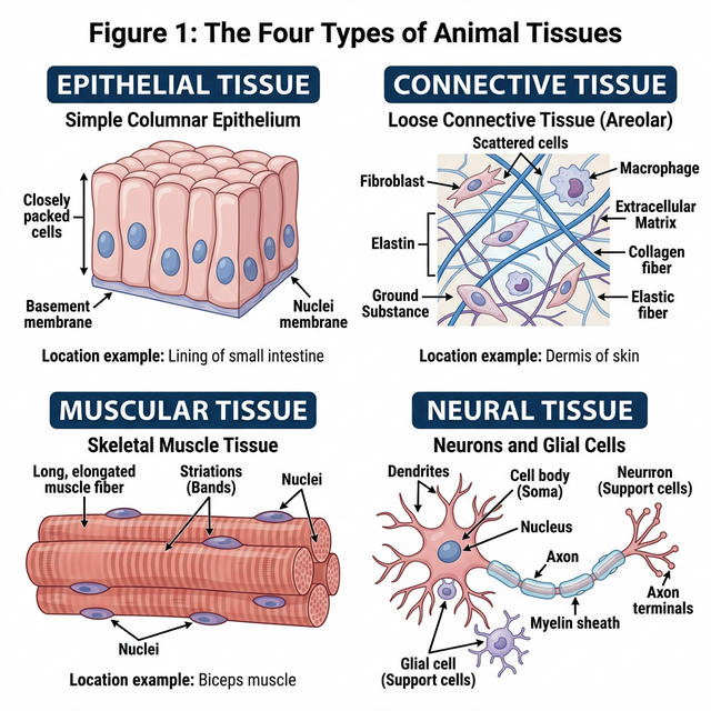
Figure 7.1: The four basic types of animal tissues
Epithelial Tissue
We commonly refer to an epithelial tissue as epithelium (pl.: epithelia). This tissue has a free surface, which faces either a body fluid or the outside environment and thus provides a covering or a lining for some part of the body.
- Simple epithelium is composed of a single layer of cells and functions as a lining for body cavities, ducts, and tubes.
- Compound epithelium consists of two or more cell layers and has protective function as it does in our skin.
Connective Tissue
Connective tissues are most abundant and widely distributed in the body of complex animals. They are named connective tissues because of their special function of linking and supporting other tissues/organs of the body. They range from soft connective tissues to specialised types, which include cartilage, bone, adipose, and blood. In all connective tissues except blood, the cells secrete fibres of structural proteins called collagen or elastin.
Muscular Tissue
Each muscle is made of many long, cylindrical fibres arranged in parallel arrays. These fibres are composed of numerous fine fibrils, called myofibrils. Muscle fibres contract (shorten) in response to stimulation, then relax (lengthen) and return to their uncontracted state in a coordinated fashion. Muscular tissue is of three types: skeletal, smooth, and cardiac.
Neural Tissue
Neural tissue exerts the greatest control over the body’s responsiveness to changing conditions. Neurons, the unit of neural system are excitable cells. The neuroglial cell which constitute the rest of the neural system protect and support neurons.
Morphology and Anatomy of Frog
Frogs are amphibians; they can live both on land and in freshwater. The most common species of frog found in India is Rana tigrina. They do not have constant body temperature; their body temperature varies with the temperature of the environment (poikilotherms or cold-blooded).
- Morphology: The frog body is divisible into head and trunk. A neck and tail are absent. The skin is smooth and slippery due to the presence of mucus.
- Digestive System: Consists of alimentary canal and digestive glands. The alimentary canal is short because frogs are carnivores and hence the length of intestine is reduced.
- Respiratory System: Frogs respire on land and in the water by two different methods. In water, skin acts as aquatic respiratory organ (cutaneous respiration). On land, the buccal cavity, skin and lungs act as the respiratory organs.
- Circulatory System: Frogs have a closed type circulatory system. They have a lymphatic system also. The heart is a muscular structure situated in the upper part of the body cavity. It has three chambers, two atria and one ventricle.
Competency Based Questions (Previous Years & Sample Papers)
Q1. An athlete sprains their ankle running on uneven terrain. The doctor diagnoses a tear in a specific dense regular connective tissue that connects muscle to bone. What is the name of this specific tissue? If the tensile strength $T$ of this tissue scales with its cross-sectional area $A$ such that $T = kA$, and the healing process initially deposits unorganized scar tissue with half the strength constant ($k_{scar} = k/2$), what must the relative area of the scar tissue ($A_{scar}/A$) be to temporarily restore the original tensile strength?
Answer
Tissue Identification: The specific dense regular connective tissue that connects skeletal muscle to bone is a tendon. (Ligaments connect bone to bone).
Mathematical Calculation: Original tensile strength: $T = kA$ New tensile strength with scar tissue: $T_{new} = k_{scar} \cdot A_{scar}$
To restore the original strength, set $T_{new}$ equal to $T$: $k_{scar} \cdot A_{scar} = kA$
Substitute the given value $k_{scar} = k/2$: $\left(\frac{k}{2}\right) \cdot A_{scar} = kA$
Divide both sides by $k$: $\frac{1}{2} A_{scar} = A$
Multiply by 2: $A_{scar} = 2A$
Therefore, the relative area of the scar tissue must be: $$ \frac{A_{scar}}{A} = 2 $$
This means the healing body must deposit twice as much cross-sectional area of unorganized scar tissue to temporarily equal the strength of the original, highly organized tendon tissue.
Q2. During a dissection of a frog (Rana tigrina), a biology student notices that the ventricle of the heart does not have a complete physical septum dividing the left and right sides. Consequently, oxygenated blood from the left atrium and deoxygenated blood from the right atrium mix before being pumped to the body. Given a scenario where the oxygen saturation of blood entering the left atrium is $96%$ and the entering the right atrium is $40%$, calculate the theoretical average oxygen saturation of the mixed blood entering the systemic circulation if the stroke volume contributions from both atria are mathematically equal ($1:1$).
Answer
If the stroke volume contributions from both atria are mathematically equal (a $1:1$ ratio), the resulting oxygen saturation of the mixed blood in the single ventricle will be the arithmetic mean of the two incoming saturations.
Oxygen saturation from left atrium ($S_{LA}$) = $96%$ Oxygen saturation from right atrium ($S_{RA}$) = $40%$
Theoretical mixed saturation ($S_{mixed}$): $$ S_{mixed} = \frac{S_{LA} + S_{RA}}{2} $$ $$ S_{mixed} = \frac{96% + 40%}{2} $$ $$ S_{mixed} = \frac{136%}{2} $$ $$ S_{mixed} = 68% $$
The theoretical average oxygen saturation of the mixed blood entering the systemic circulation would be $68%$. Note: Biologically, the frog heart has specialized structures like the spiral valve in the conus arteriosus that help reduce complete mixing, directing more oxygenated blood to the brain, but mathematically in a perfect mixing scenario, it is $68%$.
Q3. Epithelial tissues are classified based on the number of cell layers and the shape of the cells. Compare simple squamous epithelium with stratified squamous epithelium. Why would the inner lining of the human cheek be composed of stratified squamous epithelium instead of simple squamous epithelium?
Answer
Comparison:
- Simple squamous epithelium: Consists of a single layer of flat, tile-like cells resting on a basement membrane. Because it is extremely thin, its primary biological function is to facilitate rapid diffusion, filtration, or exchange of substances (e.g., in the alveoli of lungs, lining of blood vessels).
- Stratified squamous epithelium: Consists of multiple layers of cells, with the outermost layers being flat (squamous). Its primary biological function is protection against mechanical and chemical stress, friction, and abrasion.
Application to the inner cheek: The inner lining of the human cheek (buccal cavity) is subjected to constant mechanical friction from food processing (chewing, swallowing) and abrasion from teeth and tongue. If it were lined by simple squamous epithelium, the single delicate layer of cells would be easily torn away, exposing underlying connective tissue and capillaries, leading to bleeding and infection. Therefore, it is lined by stratified squamous epithelium; as the outermost layers of cells are sloughed off by friction, new cells generated by the basal layer constantly replace them, providing a durable protective barrier.