Chapter 19: Chemical Coordination and Integration
You have already learnt that the neural system provides a point-to-point rapid coordination among organs. The neural coordination is fast but short-lived. As the nerve fibres do not innervate all cells of the body and the cellular functions need to be continuously regulated; a special kind of coordination and integration has to be provided. This function is carried out by hormones. The neural system and the endocrine system jointly coordinate and regulate the physiological functions in the body.
Endocrine Glands and Hormones
Endocrine glands lack ducts and are hence, called ductless glands. Their secretions are called hormones. The classical definition of hormone as a chemical produced by endocrine glands and released into the blood and transported to a distantly located target organ has current scientific definition as follows: Hormones are non-nutrient chemicals which act as intercellular messengers and are produced in trace amounts.
Human Endocrine System
The endocrine glands and hormone producing diffused tissues/cells located in different parts of our body constitute the endocrine system.
- Hypothalamus: The basal part of the diencephalon. It produces releasing hormones (e.g., GnRH) and inhibiting hormones (e.g., Somatostatin) that regulate the synthesis and secretion of pituitary hormones.
- Pituitary Gland: Located in a bony cavity called sella tursica. The anterior pituitary secretes Growth Hormone (GH), Prolactin, Thyroid Stimulating Hormone (TSH), ACTH, LH, and FSH. The posterior pituitary stores and releases Oxytocin and Vasopressin (ADH).
- Pineal Gland: Secretes melatonin, which regulates the 24-hour diurnal rhythm of our body (sleep-wake cycle, body temperature).
- Thyroid Gland: Secretes thyroxine ($T_4$) and triiodothyronine ($T_3$), which regulate the basal metabolic rate, and Thyrocalcitonin (TCT) which regulates blood calcium levels.
- Parathyroid Gland: Secretes Parathyroid hormone (PTH), which increases the $Ca^{2+}$ levels in the blood.
- Adrenal Gland: The adrenal medulla secretes epinephrine and norepinephrine (fight or flight hormones). The adrenal cortex secretes glucocorticoids (e.g., cortisol) and mineralocorticoids (e.g., aldosterone).
- Pancreas: A composite gland. The endocrine portion (Islets of Langerhans) secretes glucagon (from $\alpha$-cells) and insulin (from $\beta$-cells) to regulate blood glucose homeostasis.
- Gonads: Testes secrete androgens (testosterone). Ovaries secrete estrogens and progesterone.
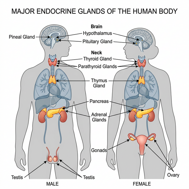
Figure 19.1: Location of major endocrine glands in the human body
Hypo- and Hyperactivity Disorders
The delicate balance of hormones is crucial. Over-secretion or under-secretion leads to severe clinical disorders.
- Dwarfism: Hyposecretion of Growth Hormone (GH) during childhood causes stunted growth.
- Acromegaly: Hypersecretion of GH in adults causes severe disfigurement, especially of the face.
- Cretinism: Hypothyroidism during pregnancy causes defective development and maturation of the growing baby leading to stunted growth (cretinism) and mental retardation.
- Goitre: Enlargement of the thyroid gland due to iodine deficiency in the diet.
- Exopthalmic Goitre (Graves’ Disease): A form of hyperthyroidism characterized by enlarged thyroid gland, protrusion of the eyeballs, and increased basal metabolic rate.
- Diabetes Mellitus: Caused by prolonged hyperglycemia due to deficiency or cellular resistance to Insulin.
- Addison’s Disease: Underproduction of hormones by the adrenal cortex alters carbohydrate metabolism causing acute weakness and fatigue.
Competency Based Questions (Previous Years & Sample Papers)
Q1. The regulation of blood glucose is tightly controlled by a negative feedback loop primarily involving the hormones Insulin and Glucagon, secreted by the Islets of Langerhans in the pancreas. Mathematically, consider normal fasting blood glucose to be set at $90 \text{ mg/dL}$. If a patient consumes a massive carbohydrate meal and their blood glucose spikes to $180 \text{ mg/dL}$, which specific cell type physically detects this mathematical error ($\Delta = +90 \text{ mg/dL}$), what hormone is immediately secreted, and what are the primary biochemical pathways activated in the liver to bring the variable back to the set-point?
Answer
Detection and Secretion: The positive mathematical deviation (hyperglycemia, $+90 \text{ mg/dL}$) is physically detected by the $\beta$-cells (beta-cells) of the Islets of Langerhans in the pancreas. In response, these cells immediately synthesize and secrete the hormone Insulin directly into the bloodstream.
Biochemical Pathways (Liver): Insulin travels to the liver (and skeletal muscles) and binds to specific tyrosine-kinase membrane receptors. This triggers a massive intracellular signaling cascade that initiates two primary biochemical pathways to reduce blood glucose:
- Glycogenesis: The rapid conversion of the excess circulating glucose into Glycogen, a highly branched, insoluble storage polysaccharide.
- Inhibition of Gluconeogenesis & Glycogenolysis: Insulin strongly inhibits the breakdown of existing glycogen and halts the de novo synthesis of new glucose from non-carbohydrate sources (amino acids/fats). By rapidly pulling glucose out of the blood and trapping it inside the liver as glycogen, the external concentration mathematically drops back towards the $90 \text{ mg/dL}$ set-point, turning off the $\beta$-cell secretion (negative feedback).
Q2. The mechanism of hormone action fundamentally differs based on the chemical nature of the hormone. Steroid hormones (like Cortisol or Estrogen) and Peptide hormones (like Insulin or Oxytocin) interact with their target cells through entirely different physical and mathematical models. Explain structurally why a steroid hormone can directly physically enter a target cell to alter gene expression, while a peptide hormone must absolutely bind to an extracellular membrane receptor, and describe the mathematical “amplification” consequence of the second messenger system used by peptide hormones.
Answer
Structural Mechanism:
- Steroid Hormones: Steroids are strictly lipid-soluble (lipophilic) molecules derived from cholesterol. Because the cell plasma membrane is a lipid bilayer, steroid hormones can freely diffuse straight through the cell membrane directly into the cytoplasm or nucleus. There, they bind to intracellular receptors to form a complex that directly binds to DNA, physically acting as a transcription factor to alter gene expression and synthesize new proteins.
- Peptide Hormones: Peptide/Protein hormones are massive, water-soluble (hydrophilic) molecules. Because they are lipophobic, they physically cannot cross the lipid bilayer. Therefore, they must act as a “first messenger” by binding exclusively to an extracellular receptor embedded on the outside surface of the target cell membrane.
Mathematical Amplification: When a single peptide hormone molecule binds to the exterior receptor, it activates a G-protein which subsequently activates an enzyme (like Adenylate Cyclase). This single enzyme physically generates thousands of “second messenger” molecules (like cyclic AMP, cAMP) inside the cell. Each cAMP molecule activates a massive cascade of protein kinases. Mathematically, this generates an enormous signal amplification cascade. A single, tiny physiological concentration of hormone ($10^{-9}$ to $10^{-12}$ Molar) mathematically results in millions of phosphorylated enzymes executing the cellular response almost instantly, making peptide hormones exceptionally potent and rapid.
Q3. The Hypothalamus is often referred to as the “Master Control Center” because it directly regulates the Pituitary Gland (the “Master Gland”). Interestingly, the hypothalamus controls the Anterior Pituitary and the Posterior Pituitary through two structurally distinct mechanisms. Describe these two separate mechanisms. For example, if a dehydrated individual needs Anti-Diuretic Hormone (ADH) to save water, exactly where is the hormone synthesized, and physically how does it reach the bloodstream?
Answer
Mechanism 1: Anterior Pituitary (Vascular Control) The hypothalamus controls the anterior pituitary entirely via a massive capillary network called the hypophyseal portal system. The hypothalamus secretes releasing or inhibiting hormones (e.g., GnRH) directly into the portal blood, which chemically travel down the stalk to mathematically stimulate or inhibit the actual endocrine cells in the anterior pituitary to synthesize and release their own distinct hormones (e.g., LH, FSH).
Mechanism 2: Posterior Pituitary (Neural Control) The posterior pituitary is not a true glandular organ; it is a direct physical, neural extension of the hypothalamus itself. The cell bodies of the neurosecretory neurons are physically located high up in the hypothalamus, where the actual hormones (ADH and Oxytocin) are synthesized.
Example of ADH: When the individual is dehydrated, osmoreceptors in the hypothalamus fire. The ADH is mathematically synthesized purely in the cell bodies within the hypothalamus. It is then physically packaged into vesicles and travels all the way down the long axons through the infundibulum stalk. The vesicles are simply stored in the synaptic knobs located in the posterior pituitary. When an action potential fires down those specific tracts, the ADH is directly exocytosed from the axon terminals into the surrounding capillaries (bloodstream). The posterior pituitary synthesizes nothing itself.