Chapter 18: Neural Control and Coordination
As you know, the functions of the organs/organ systems in our body must be coordinated to maintain homeostasis. Coordination is the process through which two or more organs interact and complement the functions of one another. For example, when we do physical exercises, the energy demand is increased for maintaining an increased muscular activity. The supply of oxygen is also increased. The increased supply of oxygen necessitates an increase in the rate of respiration, heart beat and increased blood flow via blood vessels.
In our body the neural system and the endocrine system jointly coordinate and integrate all the activities of the organs so that they function in a synchronised fashion.
Neural System
The neural system of all animals is composed of highly specialised cells called neurons which can detect, receive and transmit different kinds of stimuli. The human neural system is divided into two parts:
- Central neural system (CNS): Includes the brain and the spinal cord and is the site of information processing and control.
- Peripheral neural system (PNS): Comprises of all the nerves of the body associated with the CNS. It is divided into the Somatic neural system (relays impulses from CNS to skeletal muscles) and Autonomic neural system (relays impulses from CNS to involuntary organs and smooth muscles).
Neuron as Structural and Functional Unit
A neuron is a microscopic structure composed of three major parts, namely, cell body, dendrites and axon.
- Cell body: Contains cytoplasm with typical cell organelles and certain granular bodies called Nissl’s granules.
- Dendrites: Short fibres which branch repeatedly and project out of the cell body. They transmit impulses towards the cell body.
- Axon: A long fibre. Its distal end is branched. Each branch terminates as a bulb-like structure called synaptic knob which possesses synaptic vesicles containing chemicals called neurotransmitters. The axons transmit nerve impulses away from the cell body to a synapse or to a neuro-muscular junction.
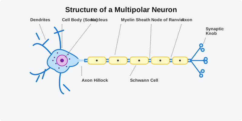
Figure 18.1: Structure of a Neuron
Generation and Conduction of Nerve Impulse
Neurons are excitable cells because their membranes are in a polarised state. Different types of ion channels are present on the neural membrane. These ion channels are selectively permeable to different ions.
- Resting Potential: When a neuron is not conducting any impulse, i.e., resting, the axonal membrane is comparatively more permeable to potassium ions ($K^+$) and nearly impermeable to sodium ions ($Na^+$). This ionic gradient is maintained by the active transport of ions by the sodium-potassium pump which transports $3 Na^+$ outwards for $2 K^+$ into the cell. As a result, the outer surface carries a positive charge while its inner surface becomes negatively charged.
- Action Potential: When a stimulus is applied, the membrane becomes freely permeable to $Na^+$. This leads to a rapid influx of $Na^+$, reversing the polarity (depolarisation). The electrical potential difference across the plasma membrane at that site is called the action potential, which is in fact termed as a nerve impulse.
Synapse
A nerve impulse is transmitted from one neuron to another through junctions called synapses. A synapse is formed by the membranes of a pre-synaptic neuron and a post-synaptic neuron, which may or may not be separated by a gap called synaptic cleft.
At a chemical synapse, when an impulse arrives at the axon terminal, it stimulates the movement of the synaptic vesicles towards the membrane where they fuse with the plasma membrane and release their neurotransmitters in the synaptic cleft. The released neurotransmitters bind to their specific receptors, present on the post-synaptic membrane.
Competency Based Questions (Previous Years & Sample Papers)
Q1. The resting membrane potential of a typical mammalian neuron is mathematically recorded as $-70 \text{ mV}$ inside relative to the outside. This electrochemical gradient is largely maintained by the electrogenic $Na^+/K^+$ ATPase pump. If a powerful neurotoxin completely binds and inhibits all $Na^+/K^+$ ATPase pumps in a neuron, calculate mathematically how an immediate single action potential event (depolarization and repolarization) will be visibly affected on an oscilloscope tracing in the millisecond timeframe. Explain what will happen to the resting potential and subsequent action potentials if the pump remains inhibited over a long period (minutes).
Answer
Immediate Single Action Potential: Mathematically and practically, an immediate single action potential will be completely unaffected and look exactly normal on the oscilloscope. Reason: A single action potential only requires an infinitely small fraction of the massive existing concentration gradients of $Na^+$ and $K^+$ to rush through the voltage-gated channels. Because the massive gradient still perfectly exists immediately after adding the toxin, the gates simply open and the ions flow normally. The $Na^+/K^+$ pump is extremely slow compared to voltage-gated channels and plays virtually no role in the millisecond duration of a single spike.
Long-Term Effect: The $Na^+/K^+$ pump’s function is purely restorative over the long term. If it is inhibited for minutes, the tiny amount of $Na^+$ that enters and $K^+$ that leaves during the natural “leakage” of the resting membrane (and during subsequent action potentials if stimulated) will never be pumped back to their proper sides. Consequently, the concentrations will slowly equilibrate. The resting potential will slowly drift from $-70 \text{ mV}$ towards $0 \text{ mV}$. Once the concentration gradients are abolished, the neuron becomes mathematically and physically incapable of generating any future action potentials, resulting in neural paralysis and death.
Q2. Multiple Sclerosis is a severe demyelinating disease where the patient’s own immune system systematically destroys the myelin sheath surrounding the axons of the Central Nervous System. Based on the bio-physics of saltatory conduction, mathematically and structurally explain why the destruction of the myelin sheath drastically slows down or completely halts the conduction of action potentials along the axon, causing severe neurological deficits.
Answer
Structure and Mathematics of Myelin: In a healthy myelinated neuron, the myelin sheath acts as a thick, powerful biological electrical insulator. It mathematically prevents ion leakage and drastically reduces the membrane capacitance. Action potentials do not travel continuously down the axon membrane; instead, voltage-gated channels are concentrated exclusively at the unmyelinated gaps called the Nodes of Ranvier. The electrical current mathematically “jumps” (saltatory conduction) extremely rapidly from node to node through the interior axonal fluid.
Pathology of Demyelination: When the myelin sheath is destroyed, two physical catastrophes occur:
- Massive Current Leakage: The extremely favorable insulating layer is gone, so the internal electrical current generated at a Node of Ranvier bleeds laterally out through the naked membrane.
- Lack of Re-amplification: Because the previously myelinated sections were specifically devoid of voltage-gated $Na^+$ channels, the naked membrane cannot regenerate or re-amplify the fading signal. Therefore, the electrical signal decays exponentially over distance just like a poorly insulated wire in saltwater. By the time the signal reaches the next node, it has fallen below the mathematical threshold voltage required to trigger the next action potential, causing the nerve impulse to literally die out and fail to reach the muscle or brain, causing the devastating symptoms of Multiple Sclerosis.
Q3. Organophosphate pesticides are lethal neurotoxins that irreversibly inhibit the enzyme Acetylcholinesterase centrally located in the synaptic cleft of neuromuscular junctions. If an unfortunate agricultural worker is heavily exposed to this pesticide, trace the specific biochemical sequence of events that occurs at their synapses. Physiologically, why does this exposure lead to violently sustained, paralyzing muscle spasms (tetanus) rather than muscle flaccidity?
Answer
Biochemical Sequence:
- A nerve impulse arrives at the synaptic knob, causing the normal exocytosis release of the neurotransmitter Acetylcholine (ACh) into the synaptic cleft.
- ACh binds perfectly to the nicotinic receptors on the post-synaptic muscle membrane, opening $Na^+$ channels, triggering an action potential, and causing the muscle fiber to contract.
- Normally, the enzyme Acetylcholinesterase (AChE) exists in the cleft specifically to immediately terminate the signal by rapidly breaking down ACh into acetate and choline, allowing the muscle to relax.
- Because the pesticide has irreversibly paralyzed AChE, the enzyme is completely broken.
Reason for Spasms: Since the ACh cannot be biochemically degraded, it permanently remains in the synaptic cleft, constantly and infinitely re-binding to the post-synaptic receptors. The post-synaptic muscle membrane is perpetually depolarized, firing endless mathematical trains of action potentials. The muscle is forced into a state of continuous, maximal, and violently sustained contraction (tetanus / spastic paralysis). The victim experiences violent convulsions and usually dies from respiratory failure as the diaphragm muscle permanently spasms and physically cannot relax to exhale.