Chapter 17: Locomotion and Movement
Movement is one of the significant features of living beings. Animals and plants exhibit a wide range of movements. Streaming of protoplasm in the unicellular organisms like Amoeba is a simple form of movement. Movement of cilia, flagella and tentacles are shown by many organisms. Human beings can move limbs, jaws, eyelids, tongue, etc. Some of the movements result in a change of place or location. Such voluntary movements are called locomotion.
Types of Movement
Cells of the human body exhibit three main types of movements, namely:
- Amoeboid: Exhibited by macrophages and leucocytes in blood, caused by streaming of protoplasm to form pseudopodia.
- Ciliary: Occurs in most of our internal tubular organs which are lined by ciliated epithelium (e.g., removing dust in the trachea, passage of ova through the female reproductive tract).
- Muscular: Movement of our jaws, limbs, tongue, etc. requires muscular movement. The contractile property of muscles is effectively used for locomotion.
Muscle
Muscle is a specialised tissue of mesodermal origin. Based on their location, three types of muscles are identified: Skeletal, Visceral, and Cardiac. Skeletal muscles are closely associated with the skeletal components of the body. They have a striped appearance under the microscope and hence are called striated muscles. As their activities are under the voluntary control of the nervous system, they are known as voluntary muscles too.
Structure of Contractile Proteins
Each skeletal muscle consists of many muscle bundles (fascicles), which in turn contain muscle fibres. Each muscle fibre contains parallelly arranged myofibrils. Each myofibril has alternate dark and light bands on it. This striated appearance is due to the distribution pattern of two important proteins – Actin and Myosin.
- Actin: Forms the light bands (I-band or Isotropic band). The filaments are thinner.
- Myosin: Forms the dark band (A-band or Anisotropic band). The filaments are thicker.
The portion of the myofibril between two successive ‘Z’ lines is considered as the functional unit of contraction and is called a sarcomere.
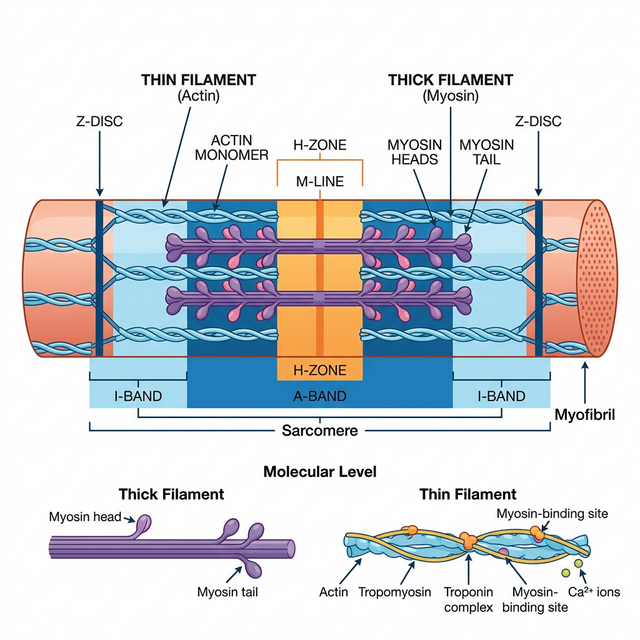
Figure 17.1: Diagrammatic representation of a Sarcomere
Mechanism of Muscle Contraction
Mechanism of muscle contraction is best explained by the sliding filament theory which states that contraction of a muscle fibre takes place by the sliding of the thin filaments (actin) over the thick filaments (myosin).
- Muscle contraction is initiated by a signal sent by the central nervous system (CNS) via a motor neuron.
- This releases a neurotransmitter (Acetylcholine) at the neuromuscular junction, generating an action potential in the sarcolemma.
- This triggers the release of Calcium ions ($Ca^{++}$) from the sarcoplasmic reticulum into the sarcoplasm.
- Calcium binds to troponin on actin filaments, unmasking the active sites for myosin.
- Utilising the energy from ATP hydrolysis, the myosin head binds to the exposed active sites on actin to form a cross-bridge, pulling the attached actin filaments towards the centre of the ‘A’ band.
Skeletal System and Joints
Skeletal system consists of a framework of bones and a few cartilages (total 206 bones in humans). Joints are points of contact between bones, or between bones and cartilages.
- Fibrous joints: Do not allow any movement (e.g., flat skull bones fusing via sutures).
- Cartilaginous joints: The bones involved are joined together with the help of cartilages, permitting limited movement (e.g., between vertebrae).
- Synovial joints: Characterised by the presence of a fluid-filled synovial cavity between the articulating surfaces. Allow considerable movement (e.g., Ball and socket joint, Hinge joint).
Competency Based Questions (Previous Years & Sample Papers)
Q1. According to the sliding filament theory, during maximum skeletal muscle contraction, the length of the A-band mathematically remains strictly constant, while the I-band shortens. If the resting length of a single sarcomere is $2.5 \mu m$, the A-band (thick filaments) is $1.6 \mu m$ long, and the specific Z-lines are assumed dimensionless, calculate the total length of the I-band region belonging to this sarcomere at rest. If the maximum contraction allows the thin filaments to meet exactly in the center of the H-zone (reducing the H-zone to $0 \mu m$), calculate the new mathematical length of the entire deformed sarcomere.
Answer
Mathematical Calculation at Rest: Length of Sarcomere ($L_{sarc}$) = Length of A-band ($L_A$) + Length of I-band components within one sarcomere ($L_I$) Given: $L_{sarc} = 2.5 \mu m$ and $L_A = 1.6 \mu m$. $$ L_{sarc} = L_A + L_I $$ $$ 2.5 \mu m = 1.6 \mu m + L_I $$ $$ L_I = 2.5 - 1.6 = \mathbf{0.9 \mu m} $$ (This $0.9 \mu m$ is split equally on both sides of the A-band, $0.45 \mu m$ per side).
Mathematical Calculation at Maximum Contraction: At maximum contraction (when the H-zone mathematically reaches $0$), the thin actin filaments (which define the I-band) are pulled entirely over the thick myosin filaments until their ends touch in the exact center. Because the A-band length ($1.6 \mu m$) remains strictly constant and the thin filaments have slid entirely over it such that there is no “non-overlapped” actin left on either side (the definition of the H-zone hitting zero and eliminating the I-band geometrically), the length of the newly contracted sarcomere is exactly equal to the length of the thick filaments (A-band). New $L_{sarc} =$ Length of A-band = $1.6 \mu m$.
Q2. Rigor mortis is the profound muscular stiffening that occurs in mammals a few hours after clinical death. It is caused by the depletion of intracellular ATP. Based directly on the biochemical steps of the cross-bridge cycle in muscle contraction, structurally explain exactly why the absence of ATP locks the muscle into a state of rigid contraction, rather than relaxation. Furthermore, what physiological event occurs roughly $48-72$ hours later that finally resolves the rigor?
Answer
Reasoning for Rigor: In the standard cross-bridge cycle, the myosin head requires the binding of a new molecule of ATP to structurally detach from the actin active site after performing the power stroke. Following death, cellular respiration rapidly ceases, and cellular ATP stores are completely depleted. Without new ATP molecules physically binding to the myosin heads, the myosin remains permanently and irreversibly locked onto the actin filaments in the contracted “cross-bridge” state. This chemical locking across billions of sarcomeres simultaneously causes the profound whole-body stiffness known as rigor mortis.
Resolution of Rigor: Rigor mortis does not resolve because the body magically synthesizes ATP or “relaxes.” It resolves $48-72$ hours later because the lysosomal membranes inside the dead cells spontaneously rupture due to decomposition. The released hydrolytic enzymes (proteases) literally digest and degrade the actin and myosin protein filaments (autolysis), mechanically severing the locked cross-bridges and softening the tissue.
Q3. Elite endurance marathon runners typically possess skeletal muscles with a massively high proportion of “Type I” (Red) muscle fibers, whereas elite Olympic weightlifters have a high proportion of “Type IIb” (White) muscle fibers. List the primary mechanisms used by Type I fibers to generate ATP, and identify the specific oxygen-binding protein causing their red color. Why does the metabolic pathway utilized by White fibers mathematically result in rapid, paralyzing muscle fatigue during sustained exercise compared to Red fibers?
Answer
Type I (Red) Fibers:
- ATP Mechanism: They primarily utilize aerobic cellular respiration (oxidative phosphorylation) within their abundant mitochondria to generate ATP.
- Color Protein: The red color is due to a very high concentration of Myoglobin, a muscle-specific oxygen-storing protein.
Fatigue in Type IIb (White) Fibers: White fibers have very few mitochondria and almost no myoglobin. To generate the massive, instantaneous force required by a weightlifter, they rely almost exclusively on anaerobic glycolysis. Mathematically and biochemically, anaerobic glycolysis is highly inefficient (yielding only $2$ ATP per glucose) and violently fast, demanding a massive flux of glucose. This rapid uncontrolled anaerobic breakdown produces excessive amounts of lactic acid ($H^+$ ions) as a byproduct. The lactic acid rapidly decreases the intracellular pH. This acidosis violently inhibits the crucial enzymes of glycolysis (like phosphofructokinase) and interferes with calcium binding to troponin, mathematically halting the muscle’s ability to contract and producing acute, paralyzing fatigue in a matter of seconds to minutes. Red fibers avoid this by fully oxidizing glucose via the Krebs cycle, producing no lactic acid.